Mole Removal in Miami
What is Mole Removal?
A mole is a very common skin growth that usually looks like a light or dark brown raised bump anywhere on your skin. They appear because of excess skin cell growth that results in a darker pigment and raised part of the skin. Moles are usually nothing to worry about but should be checked regularly at your dermatology office.
Can I Get a Mole Removed?
Sometimes, a mole grows in a way that either interferes with your regular routine (like shaving or wearing certain clothing) or you dislike the look of the mole. In those cases, a dermatologist can remove the mole using a simple procedure.
While moles are harmless in many cases, they could also indicate a serious health problem when they change in shape, size, or color over time.

You should have the mole checked by a dermatologist if any of these changes occur. Some moles could indicate skin cancer and need to be removed for that reason.
If you notice a changing or suspicious mole, report to a dermatologist as soon as you can to have it looked at. Your dermatologist might recommend further testing or mole removal surgery if skin cancer cannot be ruled out.
Mole Mapping at Greater Miami Skin and Laser Center

At Greater Miami Skin and Laser Center, we use advanced imaging technology to identify, record, and track your existing moles. Using the Foto Finder, our skilled dermatologists can perform a more thorough assessment of your skin's overall health and develop a personalized treatment regimen as needed.
How Does Mole Mapping with Foto Finder Work?
Foto Finder is a non-invasive diagnostic and imaging tool to accurately record your skin's current features and areas of potential concern.
During your mole mapping session, several images will be taken using the Foto Finder’s advanced camera technology. You will be asked to stand in various positions to allow the Foto Finder to capture images from multiple angles for analysis and growth tracking.
Once your imaging session is complete, the device will catalog detected moles based on new growths, changing sizes, or other perimeters set by your dermatologist. These findings are color-coded for easy reference, allowing our team to overview any new developments quickly.
If an area of concern is detected, the Foto Finder can produce a highly detailed dermoscopy or videoscopy image allowing your dermatologist to evaluate certain characteristics of the lesions to determine if the growth is benign or requires further investigation.
Can I Remove a Mole Myself?
Mole removal at home is not recommended. Not only is this painful, but it can also lead to severe bleeding, infection, or other complications. Visiting your dermatologist regularly and undergoing a yearly skin cancer screening is the best way to monitor moles for changes and spot a potentially cancerous growth.
How Much Does it Hurt to Get a Mole Removed?
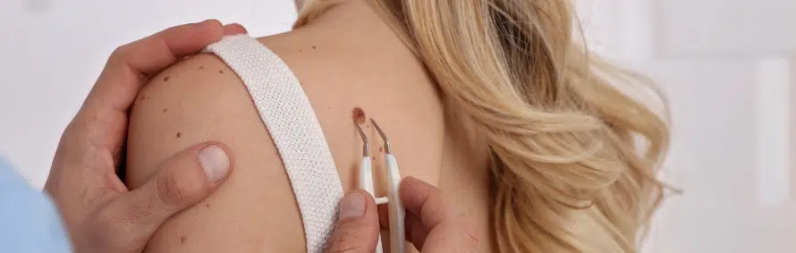
Mole removal is a relatively simple procedure involving an in-office visit to your dermatologist’s office. Your doctor will likely remove the mole by numbing the area of your skin and then cutting the mole off your skin. You may have some minor pain during or after the procedure, but overall the surgery is quick and easy to undergo. The most serious complication of mole removal surgery is infection after the procedure. Talk to your dermatologist about the steps you can take to prevent mole removal surgery infection.
How Long is Mole Removal Surgery Healing Time?
Your skin will react to mole removal surgery in a way similar to when you cut yourself on the skin’s surface. You may have some scabbing and peeling, and will likely have a scar. Most people heal from the removal after about a week, as long as you use the proper care recommended by your dermatologist.
After your mole removal surgery, you may notice the skin scabs, swells, or is red for several days following the treatment. Your dermatologist can help you prevent or minimize these effects, so be sure to ask him/her about follow-up care before leaving the office. Some dermatologists recommend keeping the area moist using vaseline to prevent swelling, soreness, redness, and other side effects.
What Does a Cancerous Mole Look Like?
The best way to accurately identify a cancerous mole is by visiting a certified dermatologist, who specializes in identifying cancerous moles. In the event that you cannot see a dermatologist right away, or if you have been instructed by your dermatologist to keep an eye on a suspicious mole over time, here are some things you can look for.
Your mole may be cancerous if:
- The mole has changed color over time
- The mole has changed shape over time
- The mole is not round/circular in shape
- The mole is asymmetrical
- The mole has grown large or protrudes excessively
- The mole has an irregular shape or color around its edges
What If I Have a Mole That Grows Back After Removal?
After a mole is removed, some of the mole skin cells could be left behind below the skin’s surface. When this happens, those skin cells can regenerate and the mole could reappear after the removal surgery. This does not always happen, but it’s difficult to predict when it will occur. The best way to remedy a mole that has grown back is by visiting your dermatologist for an evaluation of the new mole. Your doctor can examine the skin and determine what the next best step would be to prevent the regrowth in the future.
Types of Moles
Understanding the different types of moles can help you better assess any changes in your skin and determine whether a mole should be monitored or removed. Moles vary widely in shape, size, and color. Here's an overview of the primary types you might encounter:

1. Congenital Moles
Congenital moles are present at birth. These moles occur in about 1 in 100 people and can vary in size. While most congenital moles remain benign, larger ones (greater than 20 centimeters) may have a higher risk of developing into melanoma later in life. Regular monitoring by a dermatologist is recommended.
2. Acquired Moles
Acquired moles are those that develop after birth, typically appearing during childhood and adolescence. They are usually smaller than congenital moles and tend to be less than a quarter inch in diameter. Acquired moles are generally benign but should be monitored for any changes in size, color, or shape.
3. Atypical Moles (Dysplastic Moles)
Atypical moles are unusual-looking moles that can look like melanoma but are benign. They are larger than ordinary moles and have irregular and indistinct borders, and their color can vary from one area to another. People with atypical moles are at increased risk of melanoma, making regular skin checks important.
4. Spitz Moles
Spitz moles are uncommon and typically appear in children and teenagers, but they can also develop in adults. They are usually pink, raised, and dome-shaped. Although they are generally benign, their resemblance to melanoma can make diagnosis challenging, necessitating a professional evaluation for accurate identification.
5. Halo Moles
Halo moles are characterized with a white ring, or halo, around it. This occurs when the body's immune system attacks the cells within the mole. While halo moles are usually benign, any changes in the mole's appearance should be evaluated.
6. Blue Moles
Blue moles have a distinct blue or blue-gray color due to pigment deep within the skin. They are generally benign but should be observed for changes over time.
Each type of mole has unique characteristics and potential risks associated with it. Regular self-examinations and professional skin checks are crucial to monitor for any changes that might indicate a problem. If you notice a new mole or changes in an existing mole, such as asymmetry, border irregularities, color changes, diameter growth, or evolution over time, consult with a dermatologist for a comprehensive evaluation.
Will Mole Removal Leave A Scar?
Yes, mole removal can leave a scar, but the extent and visibility of the scar depend on several factors, including the mole removal technique used, the size and depth of the mole, the location on the body, and how well your skin heals.
The appearance of scars can also be minimized with proper wound care post-procedure and, if necessary, treatments such as silicone gel sheets, steroid injections, or laser treatments to improve scar appearance. Consulting with a dermatologist about the best method for mole removal and follow-up care can help reduce the likelihood of prominent scarring.